
"Everything that can be invented has been invented." This is a famous quote from the commissioner of the United States Patent and Trademark Office in early 1900. I have had almost the same idea about microscopy until MUSE (microscopy with ultraviolet surface excitation) made me believe there are still efficient and simple technologies that have not been invented yet. We owe this great methodology to a discovery by Dr. Stavros Demos, who tuned down the excitation wavelength of his laser from above 400 nm all the way to 210 nm while investigating excitation behavior of different intrinsic fluorophores in tissue. He did see those fluorophores, as expected, but more importantly he also observed a more interesting feature: the images got increasingly sharper as he moved towards shorter wavelengths, and below a certain wavelength the surface of the tissue suddenly became clearer. The idea of extending this method for rapid histology was brought up by Dr. Levenson at UC Davis, and shortly afterwards I joined the team to help make the MUSE happen.
Its development makes for an interesting story. I started with black and white image of a (previously frozen) piece of lamb kidney which I bought at a grocery store (Fig. 1). After dipping it in eosin, a very common histology dye, I recorded my first ever image montage. It was amazing and looked as if it had been taken with a scanning electron microscope. The montage was captured with a dim LED (0.5 mW) on a very slow XY stage and with a 1-megapixel camera. It took about 15 minutes to record all the small individual fields, (we can now do this in 25 s and in color), and assemble them into the montage was like putting together a puzzle. However, the final image was gorgeous, and inspired us to continue this work.

Figure 1. Image of lamb kidney stained with eosin.
We realized the great potential of this approach and worked on combining a larger sensor scientific color camera, better objectives, more powerful LEDs, smarter algorithms and alternative acquisition methodologies. Over the past few years we acquired more than 10,000 images from a wide spectrum of human, animal and plant tissues with a range of dyes and stains. The images can be truly beautiful to look at.
I had the good fortune to work with Dr. Levenson (Fig. 2) on this project, who generously took time to work in the lab and get involved in many of the experiments. The method became quite well known from his talks in scientific venues even before we could even publish the work (which turned out to be a somewhat lengthy process). In the interim, we faced repeated requests from people asking how to cite the paper which did not yet exist. We also derived huge benefits from the invaluable work of multiple volenteers in our lab. Over the past few years we have had more than 15 enthusiastic contributors from various scientific background who dedicated their time and experience to help this technology grow.

Figure 2. Farzad Fereidouni (left) and Dr. Richard Levenson (right). Advances in Optics for Biotechnology, Medicine, and Surgery. Vail, Colorado USA.
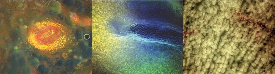
We also had our fun with Muse in the lab: for example, we imaged a slice of a hot dog, a potato and bell peppers—the hot dog was unsettling, the potato and peppers visually striking (Fig. 3).

Figure 3. Image of surface of a hot dog (left), bell pepper (middle) and sweet potato (right).
MUSE is efficient, simple and beautifully minimalistic, providing essentially a histology lab in a box. We are looking forward to seeing how it can contribute to research, clinical medicine, education at all levels, and to help provide diagnostics even in remote settings.
Our paper: Farzad, F. et al. Microscopy with ultraviolet surface excitation for rapid slide-free histology. Nat. Biomed. Eng. 1, 957–966 (2017) doi: 10.1038/s41551-017-0165-y.


Please sign in or register for FREE
If you are a registered user on Research Communities by Springer Nature, please sign in