Home to a rich microbial ecosystem, our gut digests food whilst protecting us from harmful toxins1. It also represents the largest mucosal surface area through which our immune cells interact directly with environmental antigens passing through our bodies2. Its multidimensional functions make the gut one of the most fascinating organs of the body to study, with wide-reaching implications on overall human health3.
Failure of intestinal function occurs when illness or injury of the small intestine prevents absorption of food and drink, leading to malnutrition and dehydration. A common cause of irreversible intestinal failure is “short bowel syndrome”, where large sections of the intestine have had to be surgically removed. This can occur as early as in the neonatal period, in babies born with conditions such as gastroschisis, necrotising enterocolitis or intestinal atresia.
Patients with intestinal failure require essential fluids and nutrients to be given directly into their circulating blood. In the most severe cases, patients may require transplantation. However, due to the shortage of donor organs and high complication rates, survival rates are low4.
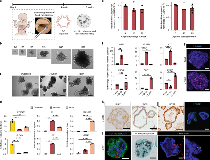
In our recent Nature Medicine paper, we first applied protocols for culturing intestinal stem cells from children with intestinal failure at Great Ormond Street Hospital, London.
We established a ‘living biobank’ of human organoids from all intestinal regions, but focused primarily on the expansion of jejunal organoids, since 90% of digestion and absorption of nutrients occur in the proximal jejunum.

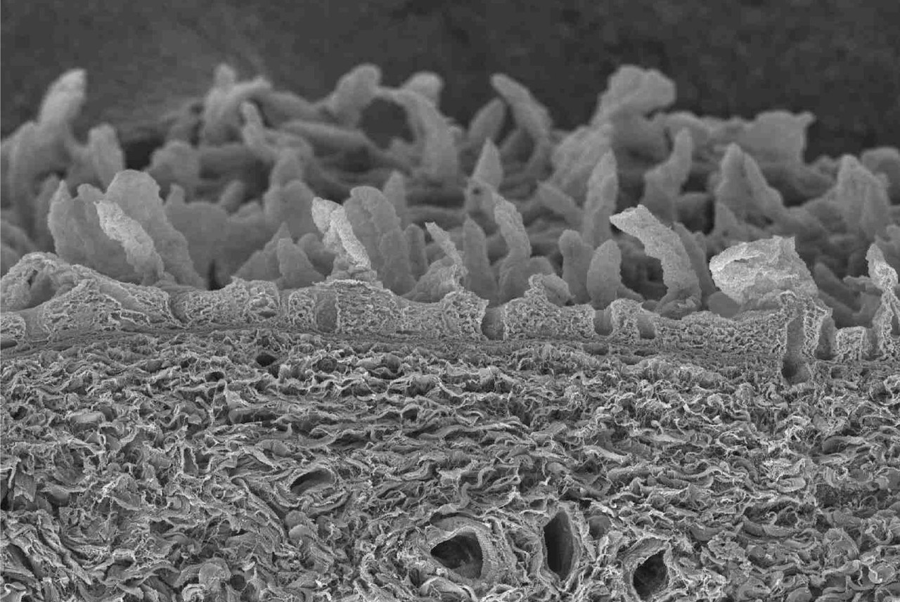
In parallel, we performed in depth characterisation of the structure and composition of decellularised intestinal scaffolds, generated from the native tissue of patients undergoing resections. Our analyses revealed the biochemical composition of small intestine and colon scaffolds to be remarkably similar. We proceeded to use both scaffolds to seed jejunal organoids, maintaining the grafts in a bioreactor culture system. We found that important features of jejunal function were recapitulated on the grafts in vitro, and the grafts survived to form luminal structures in vivo, using mouse transplantation models.
Our paper represents over 6 years of collaborative research between gastroenterologists, surgeons and basic scientists from multiple disciplinary backgrounds. The ultimate goal of our work is to restore nutritional autonomy to our patients with intestinal failure, particularly those diagnosed in early childhood. Though challenging at times, we made a concerted effort to adapt our experimental protocols in order to work with human biomaterials from our target intestinal failure patient groups wherever possible. In this way we sought to bring tissue engineering of the intestine closer to clinical translation.
We hope that our research provides a solid foundation on which our multidisciplinary team and other research groups can continue to build upon, in order to create more innovative solutions for all patients with intestinal failure.
References:
- Gebbers, J.O. & Laissue, J.A. Immunologic structures and functions of the gut. Schweizer Archiv fur Tierheilkunde 131, 221-238 (1989).
- Maloy, K.J. & Powrie, F. Intestinal homeostasis and its breakdown in inflammatory bowel disease. Nature 474, 298-306 (2011).
- Sekirov, I., Russell, S.L., Antunes, L.C. & Finlay, B.B. Gut microbiota in health and disease. Physiological reviews 90, 859-904 (2010).
- O'Keefe, S.J., et al. Short bowel syndrome and intestinal failure: consensus definitions and overview. Clin Gastroenterol Hepatol 4, 6-10 (2006).


Please sign in or register for FREE
If you are a registered user on Research Communities by Springer Nature, please sign in