
Nanoparticle therapeutics have widely been touted as the solution to solving the delivery challenges for a range of therapeutics. Nanoparticles have the potential to protect delicate cargo from degradation and also target therapeutics to specific sites of action, which maximises activity while minimising side effects. The use of lipid nanoparticle technology for the development of COVID19 vaccines has further cemented the potential of nanoparticles for therapeutic delivery.

For most applications, the therapeutic is active inside the cell, therefore delivering the nanoparticles and their cargo to the cell interior is important. The impetus for this review1 came from reading a number of papers in leading journals where cell culture studies with pharmacological inhibitors of endocytosis were used to determine the endocytic pathways of nanoparticle uptake. However, many of the conclusions from these studies didn’t seem credible. For example, we read reports of nanoparticles being internalised by caveolae in cells that lack cavins - a key structural component of caveolae. The widespread inconsistencies in the literature highlighted to us that there was a widening disconnect between the nanoparticle/materials science literature and recent developments in cell biology. We also started to wonder, are these culture studies useful? Given the ultimate goal of therapeutic nanoparticles is to treat disease, are these studies really improving efficacy in vivo?

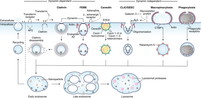
There are a number of excellent reviews on the mechanisms of endocytosis in the cell biology literature, however their focus and level of detail may not make them the ideal reference point for materials scientists. In this review, we briefly summarise the classical endocytic pathways as they are currently understood and focus on the common pitfalls with nanoparticle uptake studies. We highlight the non-specific nature of chemical endocytosis inhibitors and summarise the known off-target effects of commonly used inhibitors. We also offer a guide to genetic inhibitors of endocytosis, which offer a more targeted way to study uptake. Importantly, we summarise the endocytic mechanisms found in common human cell lines. These tables will be an important reference point when planning and analysing data from nanoparticle uptake studies.

Finally, we discuss how can these studies be improved to be more meaningful. Can tissue explants or organoids provide insights into endocytosis by differentiated cells in a more physiological setting or can cell culture systems be complemented by in vivo studies? As in vivo imaging techniques become more advanced this becomes a real possibility. We hope that this article will at least cause researchers to question the use of the standard systems used currently and look at different ways of testing the trafficking of their favourite nanoparticles.
References
1 Nature Nanotechnology 2021, 16, p 266–276
2 Nano Letters 2019, 19, p 1827–1831


Please sign in or register for FREE
If you are a registered user on Research Communities by Springer Nature, please sign in